

儿童腭裂修复术后颌面形态发育的观察研究
方成 兰东怡 张宋春媛 董晨彬
本文来源:《中华整形外科杂志》2023年7月 第39卷 第7期
DOI:10. 3760 / cma.j.cn114453-20220415-00117
作者单位:复旦大学附属儿科医院整形外科, 上海201102
通信作者:董晨彬,Email:dcb426@163.com
【摘要】
目的 探讨不同类型腭裂患儿在腭裂修复术后的颌面形态发育情况。
方法 回顾性分析 2015 年 1 月至 2020 年 12 月在复旦大学附属儿科医院整形外科行腭裂修复术的患儿及同期正常儿童的临床资料。纳入的腭裂患儿均由同一医师在 8 ~ 18 个月龄时应用松弛切口的兰氏腭裂修复术进行治疗。X 线头影测量数据和面部三维扫描资料来源于该院整形外科颌面数据库。根据腭裂类型和年龄段,将腭裂患儿分为 6 组:组 1,8 个月~1 岁Ⅰ度腭裂术前患儿;组 2,8 个月~ 1 岁Ⅱ~Ⅲ度腭裂术前患儿;组 3,2~ 3 岁Ⅰ度腭裂术后患儿;组 4,2 ~ 3 岁Ⅱ~ Ⅲ度腭裂术后患儿;组 5,4 ~6 岁Ⅰ度腭裂术后患儿;组 6,4 ~ 6 岁Ⅱ~ Ⅲ度腭裂术后患儿。根据年龄段,将正常儿童分为 3 组:组 7, 8 个月~1 岁正常儿童;组 8,2~ 3 岁正常儿童;组 9,4~ 6 岁正常儿童。多组间正态分布计量资料的比较采用方差分析,Ⅰ度腭裂、Ⅱ~Ⅲ度腭裂患儿分别与同龄正常儿童颌面特征数据比较时采用 Dunnett-t 检验。
结果 共纳入腭裂患儿 183 例,女 84 例,男 99 例,年龄(3. 6±2. 1)岁;正常儿童302 例, 女 114 例,男 188 例,年龄(4. 1±1. 9)岁。组 1~组 9 分别纳入 23、46、19、37、23、35、83、105、114 例儿童。分析结果显示,在Ⅰ度、Ⅱ~Ⅲ度腭裂患儿术前 X 线头影测量和面部三维扫描测量结果中,上颌和下颌水平长度、垂直高度和角度与同龄正常儿童相比无明显差异,而术后 2 ~ 3 岁、4 ~6 岁时的上颌和下颌水平长度、垂直高度和角度与同龄正常儿童有明显差异,且Ⅱ~ Ⅲ度腭裂患儿颌面特征数据与同龄正常儿童的差异比Ⅰ度腭裂患儿更显著。
结论 不同类型腭裂患儿在修复术后不同年龄段均出现颌面形态发育抑制,表现为上颌和下颌水平长度、垂直高度和角度的发育落后,且Ⅱ~Ⅲ度腭裂患儿术后颌面形态发育抑制较Ⅰ度腭裂患儿严重。
【关键词】腭裂;儿童;X线头影测量;三维扫描;颌面发育
The impact of children’s cleft palate repair on maxillofacial morphological growth
Fang Cheng, Lan Dongyi, Zhangsong Chunyuan, Dong Chenbin
Department of Plastic Surgery, Children’s Hospital of Fudan University, Shanghai 201102, China
Corresponding author: Dong Chenbin, Email: dcb426@163.com
【Abstract】
Objective To investigate the maxillofacial morphological development of children with different types of cleft palate after cleft palate repair.
Methods The clinical data of children who underwent cleft palate repair in the Department of Plastic Surgery, Children’s Hospital of Fudan University from January 2015 to December 2020 and normal children during the same period were retrospectively analyzed. All the included children were treated by the same physician at 8 to 18 months of age with loose incision and Langenbeck repair for cleft palate. X-ray cephalometric data and facial three-dimensional scanning data were obtained from the maxillofacial database of the Plastic Surgery Department of the hospital. According to the type and age of cleft palate, the children with cleft palate were divided into 6 groups: Group 1, 8 months to 1 years old children with grade Ⅰ cleft palate before operation; Group 2, 8 months to 1 year old children with grade Ⅱ-Ⅲ cleft palate before operation; Group 3, 2-3 years old children with grade Ⅰ cleft palate after operation; Group 4, 2-3 years old children with grade Ⅱ-Ⅲ cleft palate after operation; Group 5, 4-6 years old children with grade Ⅰ cleft palate after surgery; Group 6, 4-6 years old children with grade Ⅱ-Ⅲ cleft palate after operation. According to age, normal children were divided into three groups: Group 7, 8 months to 1 year old normal children; Group 8, 2-3 years old normal children; Group 9, 4-6 years old normal children. Analysis of variance was used to compare the measurement data of normal distribution between groups. Dunnett-t test was used to compare the maxillofacial features of children with grade Ⅰ cleft palate and grade Ⅱ-Ⅲ cleft palate with normal children of the same age.
Results A total of 183 children with cleft palate were included, including 84 females and 99 males, aged (3.6±2.1) years. There were 302 normal children, including 114 females and 188 males, aged (4.1±1.9) years. Groups 1 to 9 included 23, 46, 19, 37, 23, 35, 83, 105 and 114 children, respectively. The analysis result showed that the horizontal length, vertical height and angle of maxillary and mandibular were not significantly different from that of normal children of the same age in the preoperative cephalometric and three-dimensional scanning result of the children with grade Ⅰ and Ⅱ-Ⅲ cleft palate. However, the horizontal length, vertical height and angle of maxillary and mandibular were significantly different from that of normal children of the same age at 2-3 years and 4-6 years after surgery. Moreover, the difference between maxillofacial characteristics of children with grade Ⅱ-Ⅲ cleft palate and normal children of the same age is more significant than that of children with grade Ⅰ cleft palate.
Conclusion Maxillofacial morphological development is inhibited at different ages in children with different types of cleft palate after repair, which is manifested as the backward development of maxillofacial horizontal length, vertical height and angle of maxillary and mandible. Moreover, the maxillofacial morphological development inhibition was more serious in children with grade Ⅱ-Ⅲ cleft palate than in children with grade Ⅰ cleft palate.
【Key words】left palate; Child; X-ray cephalometry; Three-dimensional scanning; Maxillofacial development
Disclosure of Conflicts of Interest: The authors have no financial interest to declare in relation to the content of this article.
Ethical Approval: Ethical approval was given by the Medical Ethics Committee of Children’s Hospital of Fudan University (2020-477).
腭裂是常见的先天性颅颌面畸形,临床上分为软腭裂(Ⅰ度腭裂)、部分硬腭合并软腭裂(Ⅱ度腭裂)、单侧完全腭裂及双侧完全腭裂(Ⅲ度腭裂),手术是唯一的修复方法[1]。手术术式包括兰氏腭裂修复术(von Langenbeck palatoplasty)、Sommerlad腭帆提肌重建术、Furlow腭裂修复术及各种变式[2]。手术可以完美解决上腭裂隙问题,但术后颌面形态发育问题成为研究的重点,大部分研究认为腭裂术后会出现颌面形态发育的抑制,主要表现为面中部凹陷[3-5],但具体发生机制和影响因素尚不明确。本研究收集了我院整形外科近年来手术治疗的腭裂患儿的颌面特征数据,通过与正常儿童数据的对比,旨在研究不同类型腭裂修复术后的颌面形态发育情况,为临床工作提供参考。
资料与方法
一、资料选择
收集2015年1月至2020年12月在复旦大学附属儿科医院整形外科行腭裂修复术患儿及同期正常儿童的临床资料,进行回顾性分析。腭裂患儿纳入标准:(1)可收集到研究所需的完整颌面影像数据;(2)腭裂术后时间达1年及以上;(3)未合并颅颌面畸形综合征(如颅缝早闭综合征、Pierre Robin序列征)及其他畸形;(4)应用松弛切口的兰氏腭裂修复术进行治疗;(5)由同一医师行手术治疗;(6)均在8~18个月龄进行手术治疗;(7)年龄在8个月~6 岁。腭裂患儿排除标准:(1)因鼻饲喂养需插入胃管;(2)因不耐受检查拒绝配合。正常儿童的纳入标准:(1)可收集到研究所需的完整颌面影像数据;(2)年龄在8个月~6岁;(3)未合并颅颌面畸形和其他畸形。
本研究经复旦大学附属儿科医院伦理委员会批准(复儿伦审[2020]477号)。患儿监护人知情,并同意将资料用于本研究。
二、方法
(一)研究分组
研究对象共分为9组。其中,根据腭裂类型和年龄段,将腭裂患儿分为6组:组1,8个月~1岁Ⅰ度腭裂术前患儿;组2,8个月~1岁Ⅱ~Ⅲ度腭裂术前患儿;组3,2~3岁Ⅰ度腭裂术后患儿;组4,2~3 岁Ⅱ~Ⅲ度腭裂术后患儿;组5,4~6岁 Ⅰ度腭裂术后患儿;组6,4~6岁Ⅱ~Ⅲ度腭裂术后患儿。根据年龄段将正常儿童分为3组:组7,8个月~1岁正常儿童;组8,2~3岁正常儿童;组9, 4~6岁正常儿童。
(二)颌面特征数据的获取及测量
腭裂患儿和正常儿童的X线头影测量和三维扫描数据来源于我院整形外科颌面形态数据库。
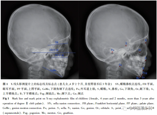
腭裂患儿和正常儿童的X线头影测量(图1):均在Healthcare Centricity RIS CE系统(美国GE公司)中进行,通过CentricityTM Universal Viewer Zero Footprint软件(版本:6. 0 SP7。0.4;美国GE公司)测量并获取颌面特征数据。所有数据按照前述分组情况逐一保存。测量数据包括反映上颌水平长度、垂直高度以及反映下颌水平长度和垂直高度的相关指标,见表1。

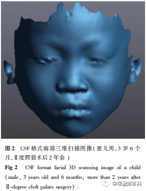
获取腭裂患儿和正常儿童面部三维扫描数据:取平卧位,完整暴露面部皮肤,利用白光三维扫描仪器(Creaform 3D Scanner,加拿大Creaform公司)对儿童面部持续进行5~7 s迅速扫描,获取CSF格式的三维图像(图2),使用VXelements 3。0软件转换输出成STL格式图像,再利用3-matic软件导入并测量颌面特征数据(图3)。所有数据按照上述分组情况逐一保存。测量数据包括反映上颌水平长度、垂直高度及反映下颌水平长度、垂直高度的相关指标,见表1。
三、统计学处理
采用SPSS 25。0统计学软件进行数据分析。符合正态分布计量资料以±s表示,相同年龄段的Ⅰ度腭裂、Ⅱ~Ⅲ度腭裂患儿与正常儿童的颌面特征数据比较,采用单因素方差分析,若差异有统计学意义,采用Dunnett-t检验进行组间两两比较。P<0。05为差异有统计学意义。
结 果
一、纳入儿童的整体情况
......
中华整形系列讲读|开播啦

14
第十四期内容
乳房再造手术时机、分期名词规范和双血管蒂腹壁
下动脉穿支皮瓣乳房再造

长按识别二维码关注视频号
观看更多精彩内容
微信视频号|中华整形外科
中华整形外科杂志
2023年 ▶▶▶

期刊官网:http://zhzxwkzz.yiigle.com
2023年每期35元,共12期,全年420元。
邮局订阅:可在全国各地邮政局订购,邮发代号80-855
网上订阅:中华医学会杂志社菁医汇商城
网址:http://jingyihui.org/shop/product/show/0/2253.html
电话:010-51322386
微信订阅:直接扫描下方二维码(手机端长按识别进入),订阅全年各期或选择性订阅某期《中华整形外科杂志》

过刊购买:中华医学会杂志社菁医汇商城,网址:http://jingyihui.org/shop/product/show/0/2253.html
目前可购买的过刊期有:目前可购买的过刊期有:2020-2022年1-12期. 电话:010-51322386


投稿方法:登录中华医学会网站http://cmaes.medline.org.cn,进行注册。注册成功后,申请成为《中华整形外科杂志》作者,即可投稿。如有问题,请致电:010-53968262。


推荐阅读
西藏高原地区唇腭裂畸形修复经验总结
唇腭裂术后继发上颌骨畸形的正颌外科治疗缺损修复中的应用
三维打印个性化托盘在唇腭裂新生儿术前鼻牙槽塑形治疗取模中的应用研究
原创文章,作者:中华整形外科,如若转载,请注明出处:https://www.meiye.net/367262.html