

面部老化中颞部软组织损失的量化研究
陈鹏飞 张恒术 杨娥 陈森
本文来源:《中华整形外科杂志》2023年2月 第39卷 第2期
DOI:10. 3760 / cma.j.cn114453-20220415-00116
作者单位:重庆医科大学附属第一医院烧伤医疗美容科, 重庆400016
通信作者:张恒术,Email:512442489@qq.com
【摘要】
目的 应用磁共振成像(MRI)方法对颞部软组织厚度行多平面量化分析,初步建立颞部软组织厚度的测量数据集,为颞部年轻化诊疗提供临床参考。
方法 采集2016年12月至2021年12月于重庆医科大学附属第一医院行MRI检查的我国西南地区汉族人群头颅高清影像进行回顾性分析。共设置12个测量点指标,较平均分布于颞部上、中、下、前、后区域,包括:颞部全层软组织厚度(NA、Na、MA、Ma、GA、Ga)及颞肌厚度(NB、Nb、MB、Mb、GB、Gb)。按性别、年龄(≤30岁、31~40岁、 41~50岁、51~60岁、>60岁)、身体质量指数(BMI,<21 kg/m2、21~26 kg/m2、>26 kg/m2)分为不同亚组,比较不同性别、年龄、BMI情况下的颞部软组织厚度的差异。正态分布计量资料2组间比较采用独立样本t检验,多组间比较采用单因素方差分析。
结果 (1)共纳入278例患者,男 140例, 女138例,年龄12~82岁,平均44.5岁,BMI 18.4~33.4 kg/m2。按年龄分组情况:≤30岁, 56例;31~40 岁,64例;41~50岁,56例;51~60岁,56例;>60岁,46例。按BMI分组情况:男性, <21 kg/m2 38例、21~26 kg/m2 56例、>26 kg/m2 46例;女性,<21 kg/m2 48例、21~26 kg/m2 50例、 >26 kg/m2 40例。(2)男性颞部软组织普遍较女性厚,以颞肌为著,6个颞肌厚度测量指标差异均有统计学意义(P<005)。(3)总体上,男性颞肌在31~40岁时及女性颞肌在≤30岁时有较其他年龄组更厚的趋势;两性群体颞肌厚度在达到高点后均随年龄增长逐渐变薄。(4)两性群体颞部全层软组织均随着BMI的增加而增厚,且下颞区全层软组织较中上颞区厚。
结论 颞部软组织厚度与性别、年龄和BMI均存在关联;两性群体颞肌随年龄增长呈逐渐变薄趋势,而颞部全层软组织随着BMI的增加而增厚。
【关键词】磁共振成像;颞部软组织;颞部凹陷;颞部填充;面部老化
Quantitative study of temporal soft tissue loss in facial aging
Chen Pengfei, Zhang Hengshu, Yang E, Chen Sen
Department of Burn and Plastic Surgery, the First Affiliated Hospital of Chongqing Medical University, Chongqing 400016, China
Corresponding author: Zhang Hengshu, Email: 512442489@qq.com
【Abstract】
Objective Magnetic resonance imaging (MRI) was used to perform multi-plane quantitative analysis of temporal soft tissue thickness, and the measurement data set of temporal soft tissue thickness was initially established to provide clinical reference for the diagnosis and treatment of temporal rejuvenation.
Methods High definition images of Han population in Southwest China who underwent head MRI in the First Affiliated Hospital of Chongqing Medical University from December 2016 to December 2021 were randomly collected in this study. A total of 12 measuring points were set, which were evenly distributed in the upper, middle, lower, anterior and posterior regions of the temporal, including the thickness of the whole temporal soft tissue (NA, Na, MA, Ma, GA, Ga) and the thickness of the temporal muscle (NB, Nb, MB, Mb, GB, Gb). They were divided into different groups according to sex, age (≤30 years old, 31-40 years old, 41-50 years old, 51-60 years old, >60 years old) and body mass index (BMI < 21 kg/m2, 21-26 kg/m2, > 26 kg/m2). The differences of temporal soft tissue thickness under different gender, age and BMI were compared. Independent sample t-test was used for comparison of normal distribution measurement data between two groups, and one-way analysis of variance was used for comparison between multiple groups.
Results (1) A total of 278 patients were included, including 140 males and 138 females, ranging in age from 12 to 82 years, with an average age of 44.5 years, and BMI of 18.4 to 33.4 kg/m2. According to age group: ≤30 years old, 56 cases; 31-40 years old, 64 cases; 41-50 years old, 56 cases; 51- 60 years old, 56 cases; >60 years old, 46 cases. According to BMI, males (< 21 kg/m2, 38 cases; 21-26 kg/m2, 56 cases; > 26 kg/m2, 46 cases) and females(< 21 kg/m2, 48 cases; 21-26 kg/m2, 50 cases; >26 kg/m2, 40 cases) were grouped. (2) Male temporal soft tissue was thicker than female, especially temporalis muscle, and the difference in 6 temporal muscle thickness measurements were statistically significant (P<0.05). (3) In general,the temporalis muscle in males at 31-40 years old and in females at ≤30 years old tend to be thicker than that in other age groups. In both sexes, the temporalis muscle became thinner with age after reaching the peak. (4) The whole layer of soft tissue in the temporal region thickened with the increase of BMI in both sexes, and the whole layer of soft tissue in the lower temporal region was thicker than that in the middle and upper temporal region.
Conclusion Temporal soft tissue thickness is associated with gender, age and BMI. The temporal muscle thickness of both sexes was gradually thinning with age, while the whole temporal soft tissue was thickening with the increase of BMI.
【Key words】Magnetic resonance imaging; Temporal soft tissue; Temporal depression; Temporal filling; Facial aging
Disclosure of Conflicts of Interest: The authors have no financial interest to declare in relation to the content of this article.
Ethical Approval: Ethical approval was given by the Medical Ethics Committee of the First Affiliated Hospital of Chongqing Medical University (2021-552).
颞部软组织可分为多个层次,由浅及深包括皮肤、皮下组织、颞浅筋膜、疏松组织、颞深筋膜浅层、颞浅脂肪垫、颞深筋膜深层、颞深脂肪垫、颞肌和骨膜[1-5]。伴随着衰老,面部软组织容量会逐渐缺失,颞部则会趋于凹陷。凹陷的颞部形态会影响整体的面部美感,特别是当眶外缘、颧弓等骨性边界显现时,更加重衰老憔悴之感[6]。临床常关注的颞部凹陷区域软组织分层复杂,内有诸多血管、神经走行,考虑到颞部软组织解剖对于颞部填充技术临床应用的重要性,本研究拟探讨颞部软组织厚度及其影响因素,为达到良好的颞部年轻化效果,补充求美者颞部容积的缺失、改善颞部软组织凹陷,重塑颞部饱满和立体的轮廓,避免发生严重并发症提供临床理论指导。
资料与方法
一、资料选择
采集2016年12月至2021年12月于重庆医科大学附属第一医院行头颅磁共振成像(magnetic resonance imaging,MRI)检查的我国西南地区汉族人群头面部高清影像进行回顾性分析。纳入标准:(1)汉族,3代及以上在国内长期居住;(2)因非颞部疾病就诊;(3)受试者和(或)家属知情同意。排除标准:(1)临床资料不全;(2)颞部手术史、外伤、畸形、肿瘤、感染及可能影响测量结果的疾病,包括颞部发育异常等严重病变;(3)1年内长期服用糖皮质激素类药物;(4)MRI图像质量欠佳,图像采集不清晰、伪影、头部歪斜者;(5)颅脑手术、创伤累及颞部。
所有受检者的临床及影像学资料来自我院影像数据库开放数据集并获受试者和(或)家属知情同意。本研究经重庆医科大学附属第一医院伦理委员会批准(2021-552)。
二、 方法
(一)研究分组
将图像样本分别按性别、年龄(≤30岁、31~40岁、 41~50岁、51~60岁、>60岁)、身体质量指数(BMI,<21 kg/m2、21~26 kg/m2、>26 kg/m2)分为不同亚组进行分析。
(二)MRI图像获取
采用德国西门子公司MAGNETOM ESSENZA 1.5T医用磁共振检测仪进行头颅MRI检查,将DICOM格式的颅脑T1加权像(T1 weighted image,T1WI)平扫或增强原始图像(层厚0.8 mm、间隔0 mm)导入离线工作站,采用Syngo.via VB20A分析软件(https:// www.caresream.GCRIS)进行手动测量,每个测量点均测量2次取平均值,以减小测量误差。
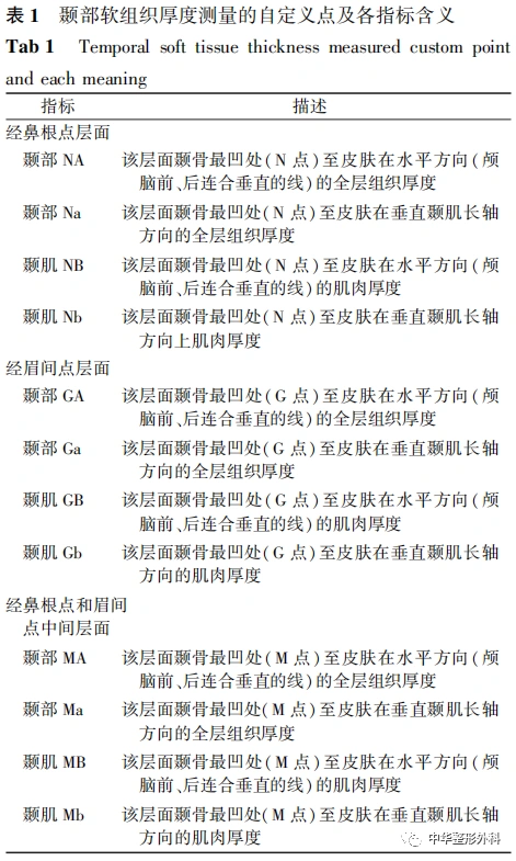
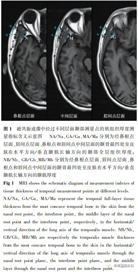
(三)颞部软组织厚度测量自定义点及各指标
根据多平面重组轴位三维T1WI平扫或增强图像,以颅脑前连合-后连合连线水平为基线[7],颞部软组织厚度测量的自定义点及12个指标含义见表1、 图1。

三、统计学处理
采用IBM SPSS Statistics 21.0统计学软件进行数据分析。正态分布计量资料以±s表示,2组间比较采用独立样本t检验,多组间比较采用单因素方差分析。P<0.05为差异有统计学意义。
结 果
一、一般资料
......
中华整形系列讲读|开播啦

10
第十期内容
重视穿支皮瓣设计进一步提升我国创面修复水平
点击链接
成为讲者
“中华整形系列讲读会”讲者报名表

长按识别二维码关注视频号
观看更多精彩内容
微信视频号|中华整形外科
中华整形外科杂志
2023年 ▶▶▶
期刊官网:http://zhzxwkzz.yiigle.com
2023年每期35元,共12期,全年420元。
邮局订阅:可在全国各地邮政局订购,邮发代号80-855
网上订阅:中华医学会杂志社菁医汇商城
网址:http://jingyihui.org/shop/product/show/0/2253.html
电话:010-51322386
微信订阅:直接扫描下方二维码(手机端长按识别进入),订阅全年各期或选择性订阅某期《中华整形外科杂志》

过刊购买:中华医学会杂志社菁医汇商城,网址:http://jingyihui.org/shop/product/show/0/2253.html
目前可购买的过刊期有:目前可购买的过刊期有:2020-2022年1-12期. 电话:010-51322386


投稿方法:登录中华医学会网站http://cmaes.medline.org.cn,进行注册。注册成功后,申请成为《中华整形外科杂志》作者,即可投稿。如有问题,请致电:010-53968262。


推荐阅读
侧颌颈靴形穿支筋膜皮瓣的应用解剖及其修复颧颞部软组织缺损的效果
额颞部扩张皮瓣修复面颈部瘢痕挛缩畸形
颞颊部除皱术结合脂肪移植在面部年轻化中的应用
原创文章,作者:中华整形外科,如若转载,请注明出处:https://www.meiye.net/180431.html