

以旋股外侧动脉斜支为蒂股前外侧皮瓣的穿支分布特点与临床应用
杨林1,2 曹阳1,2 程俊楠1,2 黄永涛1,2 柳志锦1,2 高钦锋1,2 杨成鹏1,2 孙丰文1,2 刘禹城1,2 巨积辉1,2
本文来源:《中华整形外科杂志》2023年5月 第39卷 第5期
DOI:10. 3760 / cma.j.cn114453-20230213-00031
作者单位:1苏州瑞华骨科医院手外科, 苏州215104;2苏州大学苏州医学院, 苏州215000
通信作者:巨积辉,Email:jjh2006@263.net
【摘要】
目的 探讨旋股外侧动脉斜支的穿支分布特点,以及应用以其为蒂的股前外侧皮瓣修复四肢创面的临床效果。
方法 回顾性分析2020年12月至2021年4月苏州瑞华骨科医院应用以旋股外侧动脉斜支为蒂的股前外侧皮瓣修复四肢创面的患者临床资料。术前用高频彩超于股前外侧区重点探测髂前上棘与髌骨外侧缘连线(髂髌连线)中点附近及近端粗大的旋股外侧动脉斜支穿支;根据受区缺损大小和形状,参考术前穿支定位,设计并切取以斜支为蒂的股前外侧皮瓣修复创面,供区均直接拉拢缝合。术中用钢尺测量斜支主干发出点、穿支入皮点到髂前上棘间距离;用显微标尺测量穿支口径;统计穿支数目等数据。术后对皮瓣成活及并发症情况进行观察随访,末次随访时采用综合评价量表对修复效果进行评价,90~100分为优,75~89分为良,60~74分为可,<60分为差。
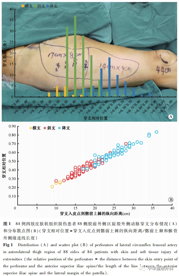
结果 共纳入84例患者,其中男62例,女22例;年龄14~82岁,平均46.9岁;手部创面32例,前臂6例,上臂3例,小腿10例,足踝部33例;创面面积为6 cm×4 cm~20 cm×45 cm。84例患者共切取88侧皮瓣(4例为双侧皮瓣),面积为7 cm×5 cm~37 cm×11 cm,其中82例85侧完全成活,1例糖尿病患者皮瓣(双侧)术后1个月完全坏死,1例34 cm的超长皮瓣远端约3 cm×3 cm发生坏死,均移植皮片修复。4例患者术后24 h内发生动脉危象,经手术探查解除动脉痉挛后皮瓣成活。供区均一期愈合。术后随访6~9个月,患者受区外形较佳,无骨髓炎等深部组织感染,皮瓣色泽、质地良好,感觉恢复为S1~S2级。综合评价量表评分为73~94分,平均88.1分,其中优33例,良46例,可5例,优良率94.0%(79/84)。88侧皮瓣术前标记215条穿支,术中实际找到穿支208条(穿支口径0.4~1.5 mm),其中斜支共发出穿支130条,88侧皮瓣均存在斜支,平均每侧1.5条,包括肌间隔穿支84条(64.6%),肌皮穿支46条(35.4%)。斜支多发自旋股外侧动脉主干,其发出后走行于股直肌与股中间肌肌间隔内,在髂髌连线中上1/3交界处分为深支与浅支。旋股外侧动脉斜支的皮肤穿支多由浅支发出,其穿支分布于髂髌连线中点及近端118条,占90.8%(118/130),在髂髌连线上0.4位点的相对位置处最集中(37条)。
结论 旋股外侧动脉斜支出现相对恒定,穿支多位于髂髌连线中点近端,且肌间隔穿支比例较高;应用以旋股外侧动脉斜支为蒂的股前外侧皮瓣修复四肢创面可获得较好的效果,在保证皮瓣血供的同时供区位置更加隐蔽。
【关键词】外科皮瓣;穿支皮瓣;股前外侧皮瓣;旋股外侧动脉斜支;穿支分布特点
基金项目:苏州市姑苏卫生人才计划项目(2020075);苏州市民生科技项目关键技术应用研究(SS202092);苏州市卫生科教临床重点病种诊疗技术专项(LCZX202026);苏州市企业工程技术研究中心专项(SZS2019263);苏州市姑苏卫生人才计划培养项目(GSWS2020116)
Distribution characteristics and clinical application of perforators of anterolateral thigh flap pedicled with oblique branch of lateral circumflex femoral artery
Yang Lin1,2, Cao Yang1,2, Cheng Junnan1,2, Huang Yongtao1,2, Liu Zhijin1,2, Gao Qinfeng1,2, Yang Chengpeng1,2, Sun Fengwen1,2, Liu Yucheng1,2, Ju Jihui1,2
1Department of Hand Surgery, Suzhou Ruihua Orthopaedic Hospital, Suzhou 215104, China; 2Suzhou Medical College of Soochow University, Suzhou 215000, China
Corresponding author: Ju Jihui, Email: jjh2006@263.net
【Abstract】
Objective To explore the distribution characteristics of the perforators of the oblique branch of the lateral circumflex femoral artery, and to report the clinical effect of the anterolateral thigh flap pedicled with the oblique branch in repairing the wounds of the extremities.
Methods The clinical data of the patients with anterolateral thigh flap pedicled with oblique branch of lateral circumflex femoral artery in Suzhou Ruihua Orthopaedic Hospital from December 2020 to April 2021 were analyzed retrospectively. High frequency color Doppler ultrasound was used to detect the large perforators of the oblique branch of the lateral circumflex femoral artery near the midpoint of the line between the anterior superior iliac spine and the lateral margin of the patella. With reference to the location of the perforators, according to the size and shape of the defect in the recipient area, the anterolateral thigh flap pedicled with oblique branch was designed and dissected to repair the wound. During the operation, the distance between the emitting point of the main oblique branch, the skin entry point of the perforators and the anterior superior iliac spine was measured with a steel ruler, the diameter of the perforators was measured with a microscale, and the number of perforators was counted. The survival and complications of the flap were observed and followed up after operation. In the last follow-up, the comprehensive evaluation scale was used to evaluate the repair effect: 90 to 100 points is excellent, 75 to 89 points is good, 60 to 74 points is average, and less than 60 points is poor.
Results A total of 84 patients were included, including 62 males and 22 females, aged from 14 to 82 years (mean 46.9 years), including 32 cases of hand wounds, 6 cases of forearm wounds, 3 cases of upper arm wounds, 10 cases of calves and 33 cases of foot and ankle wounds. The wound area was 6 cm × 4 cm-20 cm × 45 cm. A total of 88 flaps were removed in 84 patients (skin flaps on both thighs were removed in 4 patients). The size of the skin flap of 88 thighs was 7 cm × 5 cm-37 cm × 11 cm, of which 85 sides of 82 cases survived completely. One case of diabetes had complete necrosis 1 month after operation, and 1 case of 34 cm had necrosis of the distal end of 3 cm × 3 cm skin flap. Necrotic skin flaps were repaired with skin grafting. Four patients developed arterial crisis within 24 hours after operation, and those flaps survived after surgical exploration. All donor areas healed. During the follow-up of 6 to 9 months, the shape of the recipient area was normal in all patients, and there was no deep tissue infection such as osteomyelitis. The color and texture of all flaps were good. The sensation returned to S1-S2 after operation. The skin flap comprehensive evaluation scale was used to evaluate the repair effect. The patients’ score ranged from 73 to 94 points, with an average of 88.1 points. Including 33 excellent cases, 46 good cases and 5 average cases, the excellent and good rate was 94.0%(79/84). A total of 215 perforators were marked with 88 flaps before operation, and 208 perforators were found during the operation(the diameter of the perforators was 0.4-1.5 mm), of which 130 were sent out by oblique branches. There were perforators of the oblique branch in all flaps, with an average of 1.5 on each side, including 84(64.6%) septocutaneous perforators and 46(35.4%) musculocutaneous perforators. Most of the oblique branches originate from the lateral circumflex femoral artery, which runs in the intermuscular septum between the rectus femoris and the intermediate femoris muscle. It is divided into deep branches and superficial branches at the middle and upper 1/3 junction of the line between the anterior superior iliac spine and the lateral margin of the patella. The skin perforators of the oblique branch of the lateral circumflex femoral artery is mostly sent out from the superficial branch, and there are 118 perforators located at the midpoint and proximal end of the line between the anterior superior iliac spine and the lateral margin of the patella, accounting for 90.8% (118/130), reaching a peak at 0.4 (there are 37 perforators).
Conclusion The oblique branch of the lateral circumflex femoral artery is relatively constant, and most of the perforators are located near the midpoint of the line between the anterior superior iliac spine and the lateral margin of the patella, and the proportion of septocutaneous perforator is high. The distribution of perforator is regular, the blood supply is reliable, the application mode is flexible, and the donor site position is more concealed while the blood supply of the flap is secured.
【Key words】Surgical flaps; Perforator flap; Anterolateral thigh flap;Oblique branch of the lateral circumflex femoral artery; Distribution characteristics of perforator
Fund program: Health Talents Program of Suzhou Gusu District(2020075); Key Technology Application Research of Suzhou Minsheng Science and Technology Project(SS202092); Special Project on Diagnosis and Treatment Technology of Clinical Key Disease Species of Suzhou(LCZX202026); Special Project of Suzhou Enterprise Engineering Technology Research Center(SZS2019263); Suzhou Gusu Health Talent Training Project (GSWS2020116)
Disclosure of Conflicts of Interest: The authors have no financial interest to declare in relation to the content of this article.
Ethical Approval: Ethical approval was given by the Medical Ethics Committee of the Suzhou Ruihua Orthopaedic Hospital(RHGK2023024).
随着显微外科技术的发展,游离皮瓣的成活率已经显著提高,如何个性化、精确化、美观化地进行皮瓣设计,实现创面理想修复的同时减小供区的损伤成为新的研究热点[1,2]。传统股前外侧皮瓣(anterolateral thigh flap,ALTF)多以旋股外侧动脉降支发出的皮肤穿支为血管蒂,其中肌皮穿支出现概率较高,但术中解剖繁琐,对供区损伤相对较大[3],且降支穿支存在一定程度的变异,给术中切取皮瓣造成困扰。针对降支穿支缺如的情况,术中多改用其他穿支为蒂切取皮瓣,其中以旋股外侧动脉斜支报道居多,可作为ALTF的有效补充[4]。相较于降支,斜支具有肌间隔穿支比例高、供区相对隐蔽、皮瓣可切取宽度大且切取后供区可直接缝合等优点[5]。但文献报道对斜支的出现率仍存在较大争议,因此我们对以旋股外侧动脉斜支为蒂的ALTF进行了研究,通过术前进行影像学定位,术中解剖斜支主干及穿支分布特点,分析总结斜支的出现规律,以期为切取以斜支为蒂的ALTF提供一定的理论指导。
资料与方法
一、资料选择
回顾性分析2020年12月至2021年4月苏州瑞华骨科医院应用旋股外侧动脉斜支为蒂的游离ALTF修复四肢创面的患者临床资料。纳入标准:(1)外伤或感染后遗留的四肢创面;(2)采用以旋股外侧动脉斜支为蒂的ALTF修复;(3)术中术野暴露清晰,相关数据统计完整;(4)病历资料和影像学资料完整。排除标准:(1)失访或拒绝接受随访;(2)随访时间不足6个月。术前均与患者或家属签署知情同意书,本研究经苏州瑞华骨科医院伦理委员会批准(RHGK2023024)。
二、方法
(一)术前穿支定位
术前标记并连接髂前上棘和髌骨外侧缘(髂髌连线),该线为股直肌与股外侧肌肌间隔的体表投影线。在髂髌连线内外各3 cm作它的平行线,于该范围内应用高频彩超探测股前外侧区穿支血管穿出点并标记。重点探测髂髌连线中点附近及近端粗大的旋股外侧动脉斜支穿支。对于受区有主要血管缺损的患者同时行供、受区数字减影血管造影(digital subtraction angiography,DSA)。
(二)皮瓣设计
对受区行彻底清创后,根据缺损大小和形状绘制皮瓣布样。
1.设计原则:
(1)参考术前穿支定位,以髂髌连线中点近端口径较为粗大的穿支点为皮瓣的轴心点,以髂髌连线为轴心线,其内侧3 cm为皮瓣内侧切口线。(2)皮瓣的设计和切取宽度以供区能直接拉拢缝合为前提。
2.设计方法:
采用"提捏法"判断供区可供直接拉拢缝合的宽度。(1)当皮瓣布样宽度≤8 cm时,参考布样大小与形状适当放大10%~20%设计皮瓣(放大后宽度一般不超过9 cm);(2)当皮瓣布样宽度>8 cm时,采用"化宽度为长度"的方式设计分叶皮瓣;(3)对肢体超大面积皮肤及软组织缺损,单侧皮瓣不能满足创面覆盖需求,根据创面大小及形状将布样按宽度一分为二,分别设计双侧ALTF。
(三)皮瓣切取与术中数据统计
上肢创面患者采用臂丛神经阻滞加腰硬联合麻醉,下肢采用腰硬联合麻醉。患者取仰卧位,自皮瓣内侧缘切开皮肤,以电刀分离皮下组织至阔筋膜浅层并止血,遇到内侧来源的较粗大穿支先予以保留,用组织剪剪开阔筋膜,于阔筋膜下方掀起皮瓣,仔细分离皮下筋膜组织,显露股直肌与股外侧肌的肌间隔,参考术前定位标记在髂髌连线中点近端寻找斜支穿支,选择合适的目标穿支,考虑到单一穿支的供血范围不能满足皮瓣血供时,可选择多条目标穿支,若术野中未见明显粗大穿支,可适当纵向延长皮瓣切口,扩大术野寻找穿支。确认目标穿支后,切开皮瓣周缘,仔细分离皮下筋膜,逆向解剖穿支,获得足够长度血管蒂后断蒂。如果受区主干血管缺损,可切取上一级源动脉桥接受区动脉;如果受区合并空腔缺损,可携带股外侧肌填塞创面。若切取的多条穿支来源不同,不能共干时则行内增压处理。将皮瓣移植修复受区,供区皮下分层减张后直接拉拢缝合。
术中用消毒钢尺(长40 cm)测量斜支主干发出点、穿支入皮点到髂前上棘间距离,计算穿支相对位置(穿支相对位置=穿支入皮点到髂前上棘的纵向距离/髂前上棘和髌骨外侧缘连线长度);用量角器测量斜支走行角度;用显微标尺测量穿支口径;统计穿支数目等数据。
(四)术后处理及效果评价
术后常规抗凝、抗痉挛、抗感染治疗,严密观察皮瓣血运。如有血管危象,及时行血管探查修复手术。末次随访时采用综合评价量表[6],从供区、受区、患者和医生满意度等方面对修复效果进行评定,其中90~100分为优,75~89分为良,60~74分为可,<60分为差。
结 果
一、一般资料
......

中华整形系列讲读|开播啦

12
第十二期内容
不同类型支架鼻整形术的回顾性研究

长按识别二维码关注视频号
观看更多精彩内容
微信视频号|中华整形外科
中华整形外科杂志
2023年 ▶▶▶

期刊官网:http://zhzxwkzz.yiigle.com
2023年每期35元,共12期,全年420元。
邮局订阅:可在全国各地邮政局订购,邮发代号80-855
网上订阅:中华医学会杂志社菁医汇商城
网址:http://jingyihui.org/shop/product/show/0/2253.html
电话:010-51322386
微信订阅:直接扫描下方二维码(手机端长按识别进入),订阅全年各期或选择性订阅某期《中华整形外科杂志》

过刊购买:中华医学会杂志社菁医汇商城,网址:http://jingyihui.org/shop/product/show/0/2253.html
目前可购买的过刊期有:目前可购买的过刊期有:2020-2022年1-12期. 电话:010-51322386


投稿方法:登录中华医学会网站http://cmaes.medline.org.cn,进行注册。注册成功后,申请成为《中华整形外科杂志》作者,即可投稿。如有问题,请致电:010-53968262。


推荐阅读
旋股外侧动脉斜支在带蒂股前外侧皮瓣中的应用
以旋股外侧动脉横支为蒂的股前外侧皮瓣修复四肢皮肤软组织缺损
旋股外侧动脉降支游离穿支皮瓣修复伴骨外露的踝部皮肤软组织缺损
原创文章,作者:中华整形外科,如若转载,请注明出处:https://www.meiye.net/285938.html